Product Instructions for NucAP™ Pyoderma Tri-Plate (Clind/Ox)
Product Instructions for NucAP™Pyoderma Tri-Plate (Clind/Ox)
Product Overview and Intended Use
Product Overview and Intended Use
The NucAP™ Pyoderma Tri-plate (pictured below) is a next-generation rapid dual antibiotic susceptibility test for the predominant canine skin infection pathogens including S. pseudintermedius and S. aureus. The test combines the molecular power of bacterial enzymes and highly sensitive fluorescence
detection to yield results in 8 hours, much faster than the 2-5 days needed for antibiotic susceptibility testing
at off-site labs. Equipment needs include an incubator for culture plates and an inexpensive blue light box (for
information on a limited number of startup kits that include tri-plates and a suitable light box, please visit our
website: https://nptrapidtesting.com/ordering.html).
The Clind/Ox version of the NucAP™ Pyoderma Tri-plate enables susceptibility testing for oxacillin
(determines methicillin resistance) and clindamycin. This combination provides susceptibility data for first-tier
antibiotic therapies recommended for the most common form of canine pyoderma, including first-generation
cephalosporins such as cefalexin and cefadroxil (which are effective against methicillin-susceptible S.
pseudintermedius) and clindamycin (Hillier et al., 2014).
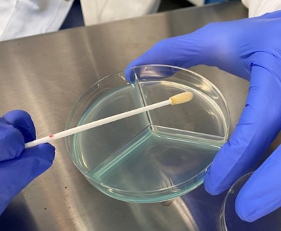
The procedural steps are as simple as inoculating the surface of the 3 chambers of the plate with a skin
swab, incubating the plate at 37 ⁰C for 8 hours to overnight, and then viewing the plate on a blue light box
. The fluorescence pattern that develops on the plate
after 8 hours indicates antibiotic resistance or susceptibility of the Gram-positive pathogens that are most
commonly found in these specimens.
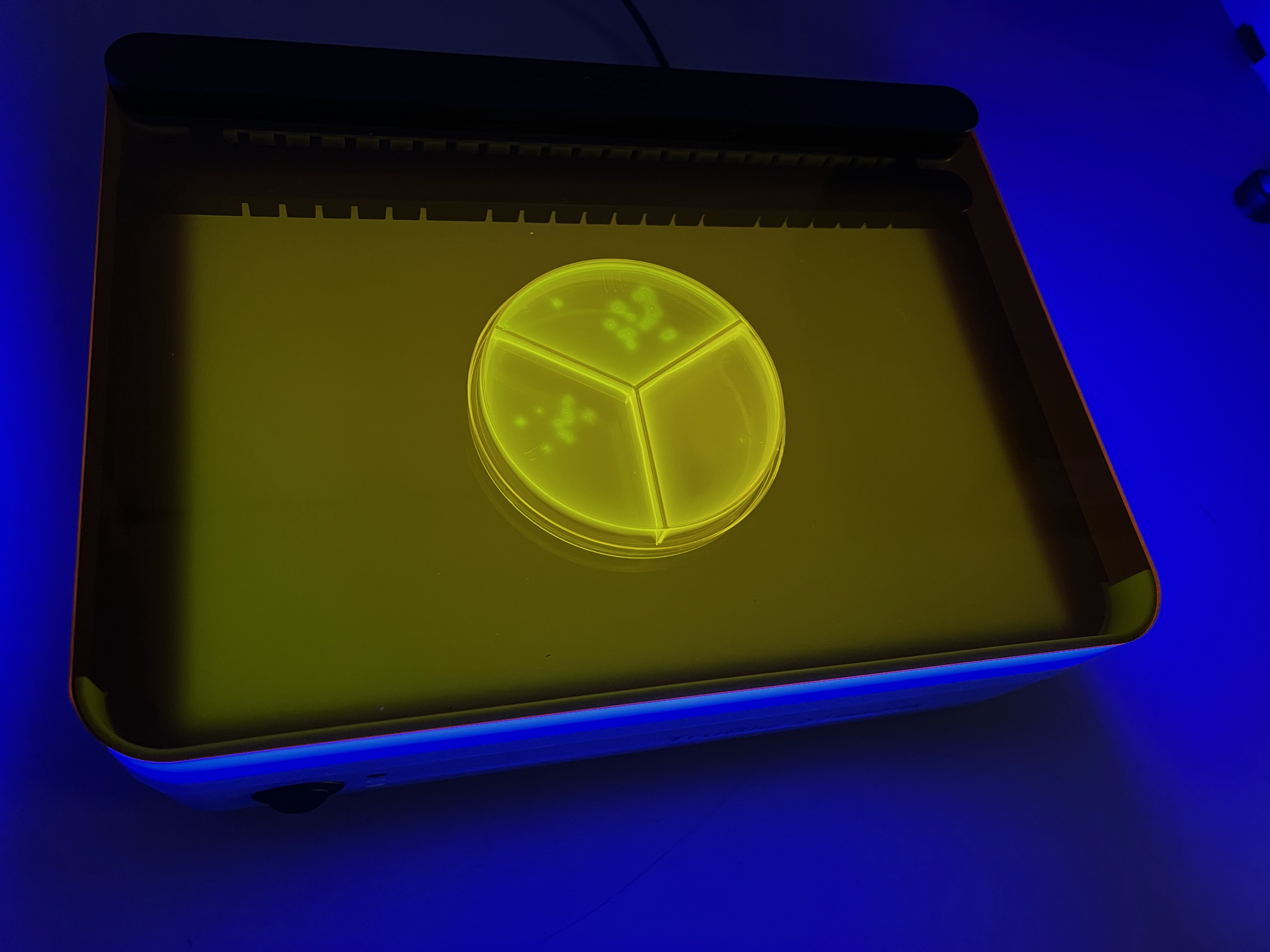
NucAP™ Pyoderma Tri-plate
Pyoderma Tri-plate on a blue light box after
8-hour incubation. (Green fluorescent spots indicate bacterial growth.)
Fluorescence development in the Gram-positive compartment indicates the presence of Gram-positive
bacteria, most likely one or more Staphylococcus species. In cases where such samples do not yield
fluorescence on the clindamycin compartment, clindamycin susceptibility is indicated. Similarly, fluorescence
development on the Gram-positive compartment, but not on the oxacillin compartment indicates methicillin
susceptibility (e.g., methicillin-susceptible S. pseudintermedius (MSSP)).
Further development of fluorescence through 19 hours of incubation can provide more information, including susceptibility data on rare slow-growing S. pseudintermedius strains and coagulase-negative Staphylococci. The NucAP™ Pyoderma Tri-plate is currently recommended for dogs with clinical lesions resembling pyoderma (pustules and papules resembling bacterial folliculitis, and epidermal collarettes) and the cytological presence of cocci within these lesions.
Safety Warnings and Precautions
Safety Warnings and Precautions
To reduce the risk of misinterpretation, users are advised to read this entire product instructions document and follow
the procedures described in the Test Procedure section below. Users are encouraged to reach out to Nuclease Probe
Technologies’ technical personnel with any questions regarding the recommended procedures and interpretation of
results. Further product instructional tutorials may be posted on Nuclease Probe Technologies’ website:
https://nptrapidtesting.com/
This product is intended for use by professional veterinarians only
Do not use this product after the expiration date printed on the package.
Do not use this product for non-veterinary applications such as diagnosing conditions in humans or for food, beverage,
water, pharmaceutical, or cosmetics testing.
Follow product storage and disposal instructions detailed below.
After inoculation, the NucAP™ Pyoderma Tri-plates may contain biohazardous microorganisms; the plates should then
be treated as biohazardous materials and handled and disposed of based on local regulations and industry standards.
NucAP™ Pyoderma Tri-plates should only be used in laboratory facilities by personnel trained in microbiological
procedures and proper handling of biohazardous materials. Users should use appropriate personal protective
equipment when handling plates, including clean gloves and lab coats
Orange-colored safety glasses such as UVEX™ S4204X Blue Light Blocking glasses are recommended to reduce
inadvertent exposure to the high intensity blue light emitted from blue light boxes
Users are advised to read and follow any safety instructions associated with accessory equipment obtained from third
parties or from Nuclease Probe Technologies. Do not operate blue light boxes without the orange screen lowered in
place; light should be switched off prior to lifting the orange screens.
This product has not been evaluated with all possible species and strains that may be encountered in veterinary
practice. The information provided here is based on in-house evaluations with canine skin lesion specimens and
bacterial strains isolated from companion animals
Do not use the plates if the media appears to be dried out or damaged, or if the plastic is cracked
This product has not been evaluated as a tool for the identification of bacterial pathogens and is not intended for such
uses.
Background
Background
Pyoderma is the number one reason for antimicrobial prescriptions in small animal veterinary practice (Hillier et al.,
2014). The absence of affordable rapid antibiotic susceptibility testing for pyoderma and the slow turnaround times of
off-site antibiotic susceptibility testing (2-5 days) have made empirical therapy for pyoderma a common approach.
However, the high and increasing rates of antimicrobial resistance among bacterial pathogens that cause pyoderma are
degrading the utility of empirical therapy.
Staphylococcus pseudintermedius, a Gram-positive, coagulase-positive bacterial species, is responsible for as much
as 92% of canine pyoderma (Lynch and Helbig, 2021). Most S. pseudintermedius strains isolated from dogs in a recent
study are resistant to one or more classes of antibiotics (Smith et al., 2020). Antibiotic susceptibility testing is thus
needed to reliably identify effective antibiotics. Recent studies report 28-33% methicillin resistance (i.e., MRSP) among
veterinary S. pseudintermedius isolates identified in New England and Texas (Little et al., 2019; Smith et al., 2020).
Canine MRSP isolates also frequently express additional resistance genes making them resistant to the antibiotics
typically used to treat MRSP, such as clindamycin, trimethoprim-sulfamethoxazole, and doxycycline (Smith et al., 2020).
Note that pathogens besides S. pseudintermedius can also cause canine pyoderma, such as Staphylococcus aureus and Staphylococcus schleiferi, but these are much less common (Hillier et al., 2014). The most common form of pyoderma in dogs is superficial bacterial folliculitis (Hillier et al., 2014).
Detailed Description
Detailed Description
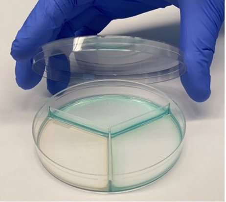
The NucAP™ Pyoderma Tri-plate includes 3 microbial growth media compartments that are selective for Gram-positive bacteria. The media in compartment I also includes oxacillin while the media in compartment II includes clindamycin. The media in all compartments includes a fluorescence indicator of enzymes that are produced by Staphylococci. Fluorescence development depends on the ability of bacteria to grow on the selective media and to produce suitable enzymes. The high sensitivity of the NucAP™ Pyoderma Tri-plate enables robust detection of Staphylococci growth on selective media prior to the appearance of visible colonies. Coagulase-positive Staphylococci, including S. pseudintermedius, produce strong enzymatic activity, resulting in visible fluorescence in as few as 6 hours. The NucAP™ Pyoderma Tri-plate thus provides a tool for determining the antibiotic susceptibility status of high-impact bacterial pathogens such as S. pseudintermedius and S. aureus much faster than current alternatives such as chromogenic agars. Rare slow-growing strains of S. pseudintermedius and S. aureus, and various Gram-positive bacterial species that are less frequent causes of canine skin infections, such as coagulase-negative Staphylococci (CoNS),will also yield fluorescence on the NucAP™ Pyoderma agar plates after overnight incubation. The NucAP™ Pyoderma Tri-plate has not been evaluated as a tool for the identification of bacterial pathogens and is not intended for such uses.
Instructions for Use
Instructions for Use
Test Procedure
Test Procedure
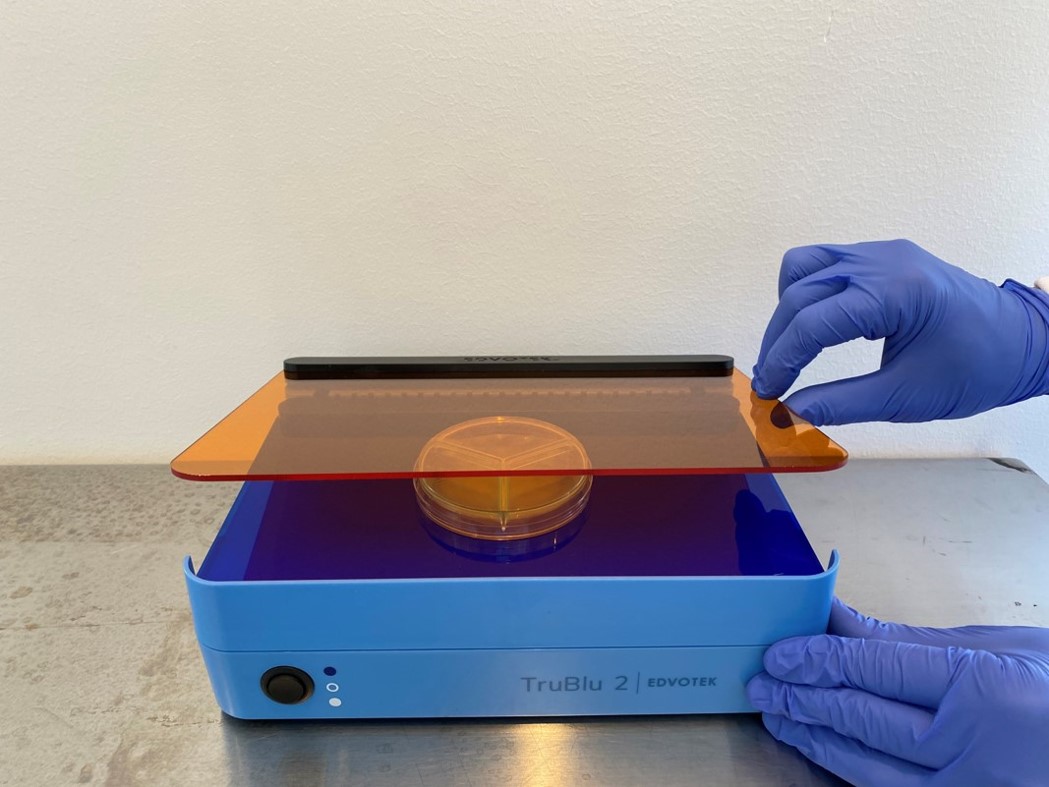
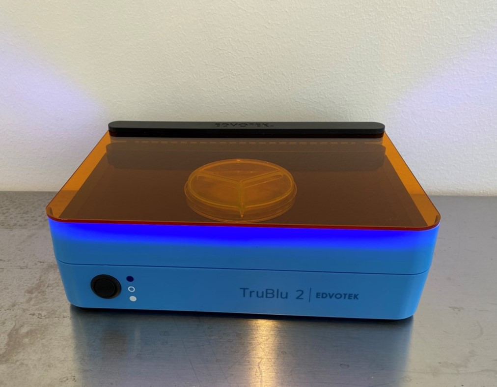
Wear clean gloves and open plates on a clean surface such as a bench-top wiped with 70% ethanol. Inoculate the media of each compartment of a fresh NucAP™ Pyoderma Tri-plate with a specimen swab. To avoid transferring antibiotics between compartments, use a different part of the swab for each compartment (i.e., rotate swab ¼ turn prior to inoculating each compartment), and inoculate the Gram-positive compartment (compartment III) first. Place the plate in a 37 ⁰C incubator for 8 hours. (When using the blue light boxes, orange-colored blue-light-blocking safety glasses are recommended as additional protection from inadvertent exposure to the high intensity light emitted from these devices.) With the light switched off, place the plate on the surface of a blue light box such as the TruBlu™ 2 Blue/White Transilluminator of Edvotek (recommended) and lower the translucent orange screen over the plate. Turn on the blue light and view the plate, noting the presence or absence of green fluorescent spots in each of the 3 compartments. If the
plate will be incubated further, limit the light exposure to ~1 minute and return plate to incubator within 5 minutes.
Turn off the light prior to lifting the orange screen and removing the plate.
Results Interpretation
Results Interpretation
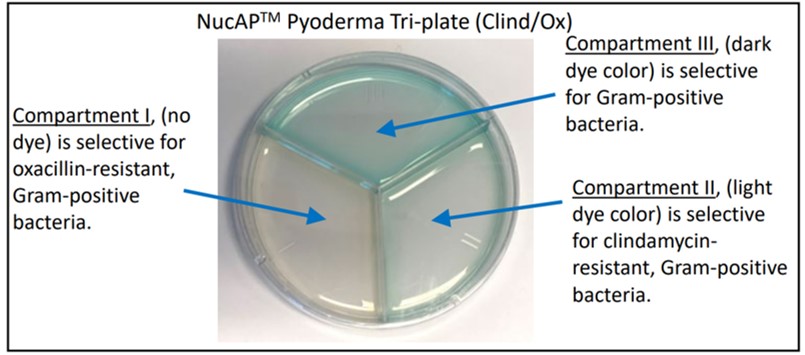
The Gram-positive compartment (indicated with number III and by the strongest dye color) serves as a positive
control for the presence and growth of bacterial pathogens that can be tested with the NucAP™ Pyoderma Tri-plate.
Green fluorescent spots visible within 6-8 hours on the Gram-positive compartment is a general indication of the
presence and growth of Gram-positive bacteria, most likely one or more Staphylococcus species. The production of
green fluorescent spots in compartments I and/or II indicates the presence of bacteria that are resistant to oxacillin
and/or clindamycin, respectively. Fluorescent spot development in compartments I and II is often delayed by about an
hour from that in compartment III. Before concluding that bacteria detected in the Gram-positive compartment are
susceptible to either oxacillin or clindamycin, users should confirm that no green fluorescent spots are developing in the
corresponding compartment I or II after 8 hours of incubation.
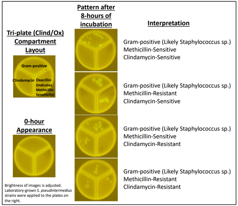
The typical fluorescence patterns generated at 8 hours is shown in the figure. The layout of the
compartments in the images is shown on the upper left image for reference. Note the absence of green fluorescent
spots on the plate image taken immediately after sample application (0-hour Appearance); this is also the typical
appearance of plates at 8 hours in which Gram-positive bacteria are not present. The plate images on the right show the
typical patterns produced by canine skin pathogens such as S. pseudintermedius and S. aureus. In all 4 cases, the
presence of green fluorescent spots in compartment III indicates the presence of Gram-positive bacteria, most likely one
or more Staphylococcus species. From top to bottom on the right, the first image shows the pattern seen with
methicillin-sensitive, clindamycin-sensitive bacteria; the second image shows the pattern seen with methicillin-resistant,
clindamycin-sensitive bacteria; the third image shows the pattern seen with methicillin-sensitive, clindamycin-resistant
bacteria; and the fourth image shows the pattern seen with methicillin-resistant, clindamycin-resistant bacteria.
Note that rare, slow-growing strains of species such as S. aureus and S. pseudintermedius can produce fluorescence
that only emerges after 7 or more hours. These strains will typically exhibit smaller green fluorescent spots at 8 hours
compared to those produced by the more common strains. Samples that yield green fluorescent spots on the Grampositive (compartment III) at 8 hours that are substantially smaller than those shown on the images below without
corresponding green fluorescent spots on compartments I and II should be incubated for an additional hour and then viewed again on a blue light box for more accurate interpretation. Finally, cases in which the initial fluorescence is only
visible after 10 hours are generally the result of coagulase-negative Staphylococci, such as S. epidermidis.
Viewing the plates on a blue light box after overnight incubation is optional, but can provide additional information.
The fluorescence pattern after 19 hours of incubation is generally more diffuse and higher in its intensity versus that
seen at 8 hours. Gram-positive bacteria of non-Staphylococcus species, such as Enterococci, may produce colonies on
the agar visible after 19 hours of incubation. These colonies generally produce limited fluorescence that is confined to
the colonies themselves, without an associated diffuse signal in the media. The presence of a colony without associated
diffuse fluorescence in the surrounding media is an indication that the bacteria are a non-Staphylococcus species.
In some cases, various types of debris present in specimen swabs can yield fluorescent spots on the plates. This
fluorescence can be distinguished from the enzyme-derived fluorescence that results from growing bacteria by its shape
and static nature as debris-based fluorescence will generally be unchanged in intensity and shape over multiple imaging periods. If this is a recurring problem, consider viewing the plates immediately after inoculations. Fluorescence visible at
this initial time-point can be assumed to be the result of debris.
Storage
Storage
Upon delivery, store NucAPTM Pyoderma Tri-plates at 4 ⁰C protected from light up until the expiration date marked on the package.
Disposal
Disposal
After application of specimens, the NucAPTM Pyoderma Tri-plates may contain biohazardous microorganisms. Disposal of NucAPTM Pyoderma Tri-plates should follow local regulations and industry standards that are currently in place for biohazardous materials.
Hillier, A., Lloyd, D.H., Weese, J.S., Blondeau, J.M., Boothe, D., Breitschwerdt, E., Guardabassi, L., Papich, M.G., Rankin, S., Turnidge, J.D., et al. (2014). Guidelines for the diagnosis and antimicrobial therapy of canine superficial bacterial folliculitis (Antimicrobial Guidelines Working Group of the International Society for Companion Animal Infectious Diseases). Vet Dermatol 25, 163-e143.
Little, S.V., Bryan, L.K., Hillhouse, A.E., Cohen, N.D., and Lawhon, S.D. (2019). Characterization of agr Groups of Staphylococcus pseudintermedius Isolates from Dogs in Texas. mSphere 4.
Lynch, S.A., and Helbig, K.J. (2021). The Complex Diseases of Staphylococcus pseudintermedius in Canines: Where to Next? Vet Sci 8.
Smith, J.T., Amador, S., McGonagle, C.J., Needle, D., Gibson, R., and Andam, C.P. (2020). Population genomics of Staphylococcus pseudintermedius in companion animals in the United States. Commun Biol 3, 282.